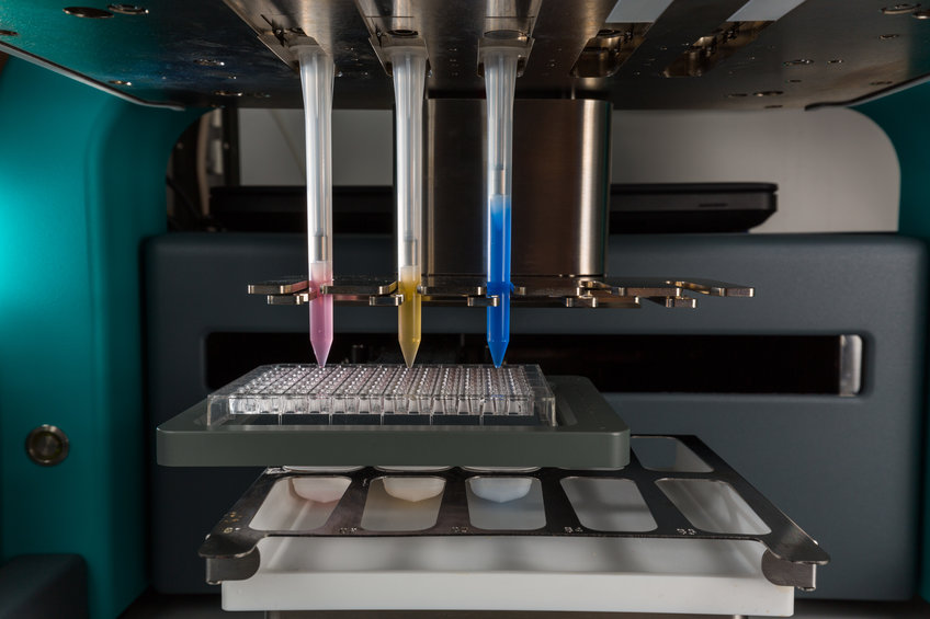
X-Ray Crystallography
The primary goal of structural biology is a mechanistic understanding of biological macromolecules and of biological processes so that they can be described in the language of physics and chemistry. This requires an exact knowledge of the three-dimensional structures of the respective molecules and of their complexes such that the spatial position of each atom can be precisely described. Since the pioneering work of Max Perutz and Sir John Kendrew X-ray crystallography has been the single most important technique for the determination of three-dimensional protein structures.
Since the pioneering work of Max Perutz and Sir John Kendrew X-ray crystallography has been the single most important technique for the determination of three-dimensional protein structures. Currently, the X-ray coordinates for more than 50,000 macromolecules can be found in the protein database (PDB), far exceeding any other comparable technique. To obtain a crystal structure of a macromolecule one needs to purify several hundred micrograms of material in soluble form and one needs to obtain crystals that can be as small as a few micrometers in diameter. These crystals are then exposed to diffract a finely tuned monochromatic X-ray beam, nowadays typically generated by a synchrotron cyclic particle accelerator such as the Swiss Light Source, where the Max Planck Society partially owns one of the beamlines (PX II). Once diffraction data are recorded, electron density throughout the crystal can be computed and macromolecular models can be built.
Equipment
- Genomic Solutions Honeybee 963 Crystallization Robot
- TTP Labtech Mosquito Crystallization Robot
- TTP Labtech Dragonfly Screen Optimisation Robot
- Dornier LTF PIRO Pipetting Robot
- Formulatrix Rock Imager 54 Imaging System @ 8 °C
- Formulatrix Rock Imager 182 Imaging System with UV optics @ 20 °C
- Xenocs sealed Tube X-ray Generator
- Oxford Cryosystems Cryostream 700
- MarResearch MAR345 Image Plate Detector
- Regular access to beamline X10SA at the SLS with PILATUS 6M Detector